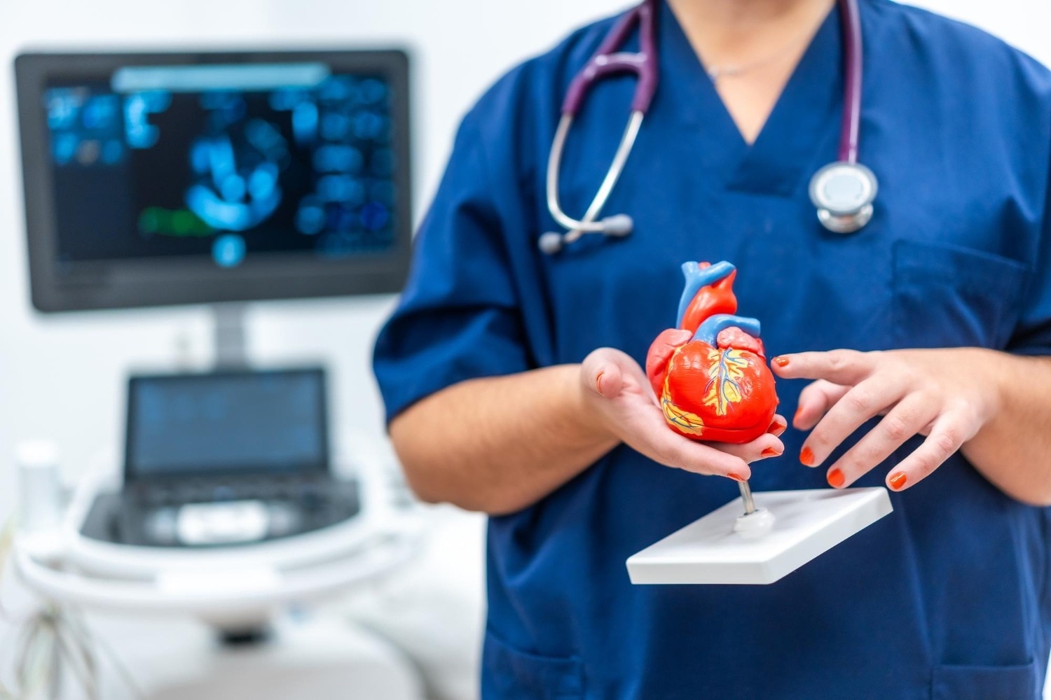

A catheter-based procedure can reduce the need for open surgery, but it is not automatically safer or suitable for every structural heart problem. By the end, you will know how doctors distinguish the condition, match anatomy and risk to a treatment, understand the testing and recovery involved, and compare long-term trade-offs.
Key takeaways
- Match treatment to the valve, defect, symptoms and imaging findings.
- Ask how echocardiography, CT, angiography and catheter tests affect the recommendation.
- Discuss recovery time, bleeding, infection, stroke and rhythm risks before treatment.
- Confirm medication duration, durability expectations and future procedure plans.
Start With the Lesion, Not the Procedure
Structural heart disease changes the heart’s physical architecture: its valves, chambers, walls, septa or nearby great vessels. It differs from coronary artery disease, which narrows blood vessels; rhythm disorders, which disturb electrical signals; and heart failure, which describes impaired pumping or filling. One person can have more than one of these conditions.
The lesion, not the procedure name, determines the treatment:
- A hole in the atrial or ventricular septum may require closure, but its size, location, surrounding tissue and shunt volume decide whether a device or surgery is safe.
- Aortic stenosis, mitral regurgitation and rheumatic mitral stenosis are different valve problems. Their leaflets, calcium, leakage, pressure effects and ventricular function determine whether repair, replacement, balloon treatment or medical care fits.
- A left-atrial appendage clot-risk problem is not a valve lesion, so left atrial appendage occlusion requires a separate assessment of stroke risk, bleeding risk and appendage anatomy.
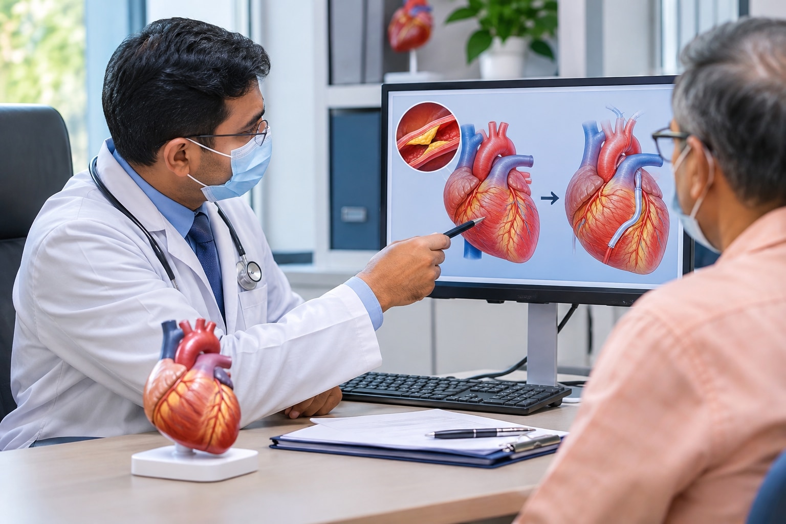
HeartDiseaseTreatment should begin with a transthoracic echocardiogram and a clinical examination, then use transesophageal echocardiography, cardiac CT or MRI when the images or procedural measurements require them. A single valve gradient or ejection-fraction number cannot select treatment by itself.
For StructuralHeartDiseaseInPune, ask whether a Heart Team will review the lesion, symptoms, ventricular response, kidney function, medicines and procedural risk together. That assessment prevents a less-invasive procedure from being chosen for anatomy it cannot safely treat.
Which Catheter-Based Options Match Your Anatomy?
A catheter procedure is suitable only when the defect’s size, location and surrounding tissue provide a safe landing zone. Your symptoms, ventricular function, surgical risk, age, life expectancy, kidney function and ability to take antithrombotic medicine also affect the choice. A Heart Team confirms eligibility using echocardiography and, when needed, transesophageal echocardiography or cardiac CT.
| Option | Best match | Eligibility signals |
|---|---|---|
| Catheter ASD or selected VSD closure | A hole between heart chambers | Significant shunt, suitable rims and no severe pulmonary hypertension; some VSDs are too close to valves or the conduction system |
| Balloon mitral commissurotomy | Rheumatic mitral stenosis | Significant symptoms, pliable valve, no left-atrial thrombus and no more-than-mild mitral regurgitation; heavy calcium or major leakage argues against it |
| TAVR | Severe aortic stenosis | Appropriate valve and artery anatomy, feasible vascular access and a risk–durability profile accepted by the Heart Team; age, life expectancy and patient preference matter |
| TEER, such as MitraClip or PASCAL | Selected severe mitral regurgitation | Suitable leaflet anatomy; high or prohibitive surgical risk for primary disease, or persistent secondary regurgitation after guideline-directed heart-failure therapy |
| Left atrial appendage occlusion | Stroke prevention in atrial fibrillation | A suitable appendage, no active thrombus and a strong reason to avoid long-term anticoagulation |
A failed anatomy match can leave residual leakage, device migration or emergency surgery as the consequence. Ask whether the centre provides CT or TEE planning, kidney and anticoagulant management, emergency backup and follow-up for valve performance, rhythm problems and residual defects; an AdvancedCardiacCare consultation should address each point.
How Tests and the Heart Team Select the Safest Route
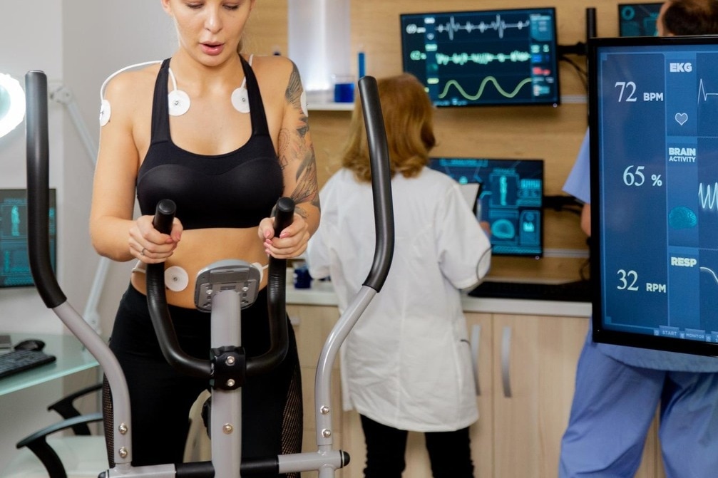
A transthoracic echocardiogram (TTE) measures valve narrowing, leakage, chamber size and ventricular pumping. Transoesophageal echocardiography (TEE) shows valve edges, clots, septal defects and suitability for a clip or balloon procedure. Cardiac CT maps calcium, the valve annulus and blood-vessel access. ECG, blood tests, kidney function, and coronary angiography add rhythm, contrast-safety and artery information.
| Route | Strengths | Trade-offs and checks |
|---|---|---|
| Catheter treatment | Smaller access route and shorter recovery for suitable anatomy | Vascular injury, stroke, contrast-related kidney injury, residual leak, pacemaker need and rare emergency surgery |
| Repair surgery | Durable repair, especially for suitable degenerative mitral regurgitation | Chest surgery, longer recovery and risks from anaesthesia, bleeding and infection |
| Replacement surgery | Treats valves unsuitable for repair or catheter therapy | Prosthesis choice, anticoagulation, recovery time and future re-intervention require planning |
The multidisciplinary Heart Team compares these routes against anatomy, symptom severity, ventricular function, age, life expectancy, surgical risk, vascular access, durability and your preferences. For TAVR, it weighs transcatheter replacement against surgical replacement; for mitral regurgitation, it favours surgery when durable repair is safe and considers TEER for suitable high-risk patients.
Balloon mitral commissurotomy requires favourable rheumatic-valve anatomy, no left-atrial thrombus and no more-than-mild regurgitation. Ask who reviews the scans, manages anticoagulants and kidney risk, provides anaesthesia and emergency backup, and follows valve performance, rhythm and residual defects. That process makes HeartDiseaseTreatment a patient-specific decision rather than a choice based on the least invasive label.
What Happens During Recovery—and What Can Go Wrong?
Before a catheter procedure, expect blood tests for kidney function and blood counts, medication instructions, fasting, and imaging such as transesophageal echocardiography or cardiac CT. Ask who will review your case, which anaesthesia is planned, and what emergency surgical backup is available.
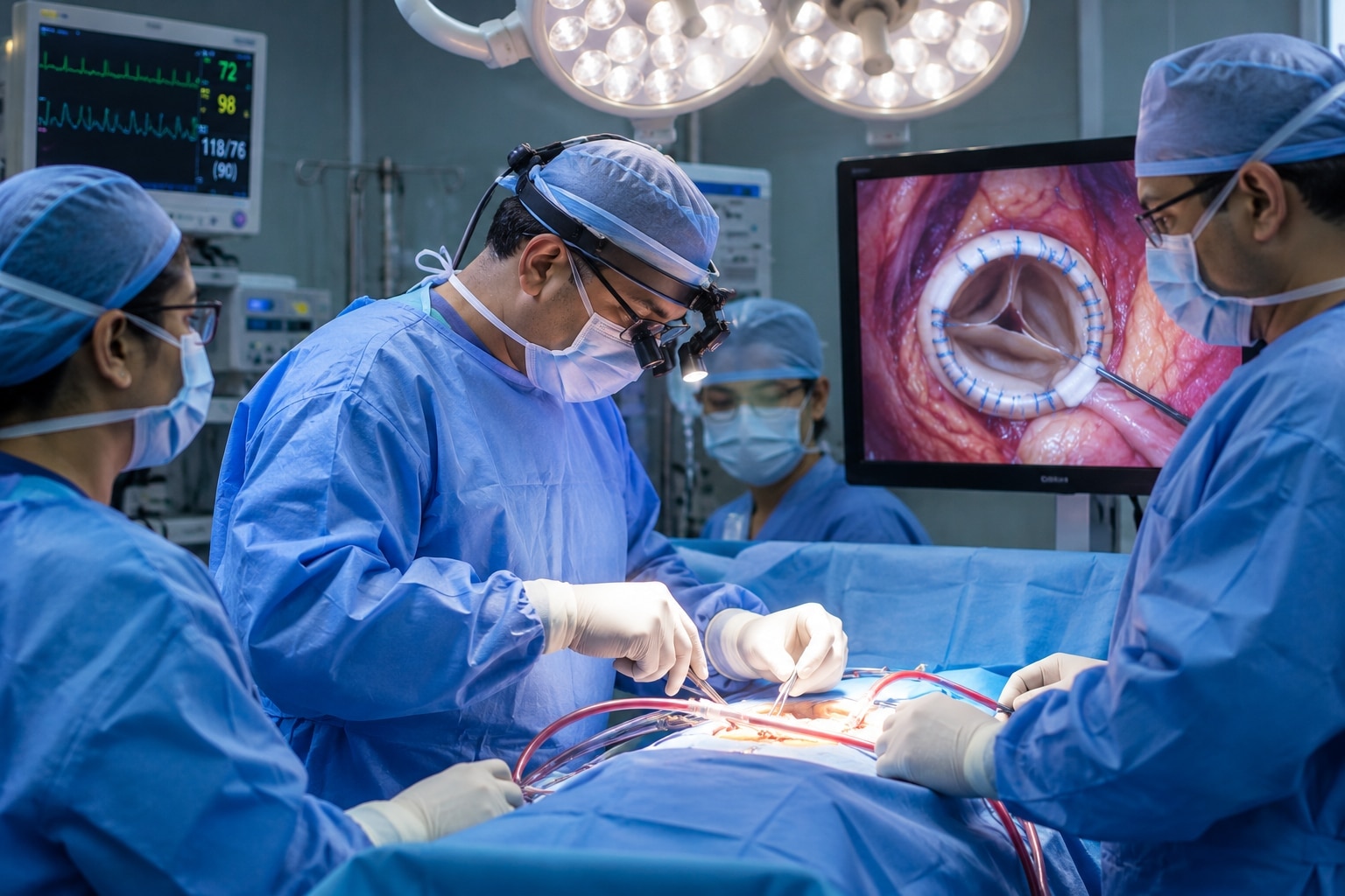
During the procedure, a catheter usually enters through the groin, guided by X-ray and echocardiography. You may receive local anaesthetic with sedation, or general anaesthesia for longer or more complex repairs.
Afterward, staff monitor the access site, rhythm, blood pressure and kidney function; you may walk later that day or the next, but follow-up imaging and medication review remain essential.
A StructuralHeartDiseaseInPune decision should weigh recovery against the hazards of both routes:
| Option | Catheter procedure | Open surgery |
|---|---|---|
| Recovery | Smaller access wound; discharge often sooner | Chest incision; longer hospital stay and activity restrictions |
| Trade-off | Less surgical trauma, but device-specific risks and possible repeat intervention | More pain and bleeding risk, but direct repair can offer greater durability for suitable anatomy |
| Serious complications | Stroke, bleeding, vessel injury, infection, contrast-related kidney injury, rhythm block needing a pacemaker, residual leak or device migration | Infection, bleeding, stroke, kidney injury, rhythm problems and complications from bypass |
Seek urgent help for chest pain, fainting, breathlessness, fever, a cold or bleeding puncture site, or a weak leg. A catheter procedure can require emergency surgery in rare cases, so “minimally invasive” never means risk-free.
Plan for Durability, Medication and Future Procedures
Device choice must include its next chapter, not just the procedure. A transcatheter aortic valve is not automatically permanent: younger patients need a durability discussion, including future valve-in-valve treatment. A second valve can be feasible, but it may leave a smaller opening and complicate access to the coronary arteries.
Ask how your age, life expectancy, anatomy and pacemaker risk affect TAVR versus surgery.
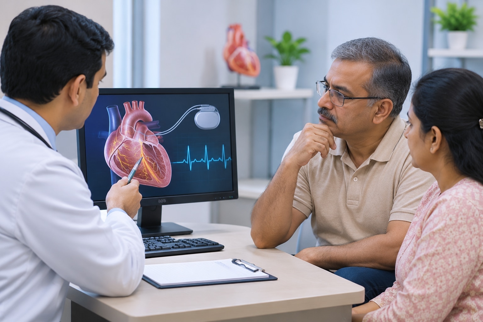
Medication plans are part of the device decision. After valve implantation or left-atrial-appendage occlusion, follow the prescribed antiplatelet or anticoagulant schedule; do not stop it for dental work or surgery without instructions. LAA occlusion is not a blanket replacement for anticoagulation, and follow-up imaging checks for device-related clot or residual leakage.
Maintain dental care, report fever promptly and ask whether you need antibiotic prophylaxis; it applies only to selected high-risk patients.
Pregnancy needs planning before intervention, especially if a mechanical valve and lifelong anticoagulation are considered. Discuss timing, medication changes and radiation exposure with cardiology and obstetrics before conception. Choose StructuralHeartDiseaseInPune care that provides local echocardiography, rhythm checks, kidney and anticoagulant review, and assessment of residual leaks.
A Cardiologist in Pune should also explain emergency backup, imaging, anaesthesia and who will manage complications—questions that make an AdvancedCardiacCare consultation clinically useful, not merely minimally invasive.
Related services
Structural Heart Disease Treatment in Erandwane, Pune Structural Heart Disease Treatment in Erandwane, Pune Structural heart diseases include congenital defects, valve disorders, and abnormalities in heart chambers. View service → |
Valve Replacement Surgery in Erandwane, Pune Valve Replacement Surgery in Erandwane, Pune Valve replacement surgery restores proper blood flow in patients with severely damaged or diseased heart valves. View service → |
Frequently asked questions
How is treatment chosen for structural heart disease?
A heart team matches the lesion and your anatomy to catheter-based treatment, surgery or monitoring after reviewing symptoms, echocardiography, CT, angiography and other tests.
Which catheter-based treatments can treat structural heart disease?
Options depend on the lesion and anatomy. They can include transcatheter valve replacement or repair, closure of selected septal defects, and catheter procedures for certain nearby vessel problems.
What tests are used before structural heart treatment?
Doctors may use transthoracic or transesophageal echocardiography, cardiac CT, cardiac MRI, coronary angiography and right-heart catheterisation to assess anatomy, pressure and procedural risk.
What should you expect during recovery after treatment?
Recovery depends on the procedure. Discuss hospital stay, activity restrictions, follow-up imaging, medication changes and warning signs such as chest pain, breathlessness, fever, bleeding or neurological symptoms.
Related service
Cardiology Consultation in Erandwane, Pune Cardiology Consultation in Erandwane, Pune A Cardiology Consultation is the first step toward understanding and managing your heart health. View service → |
Keywords