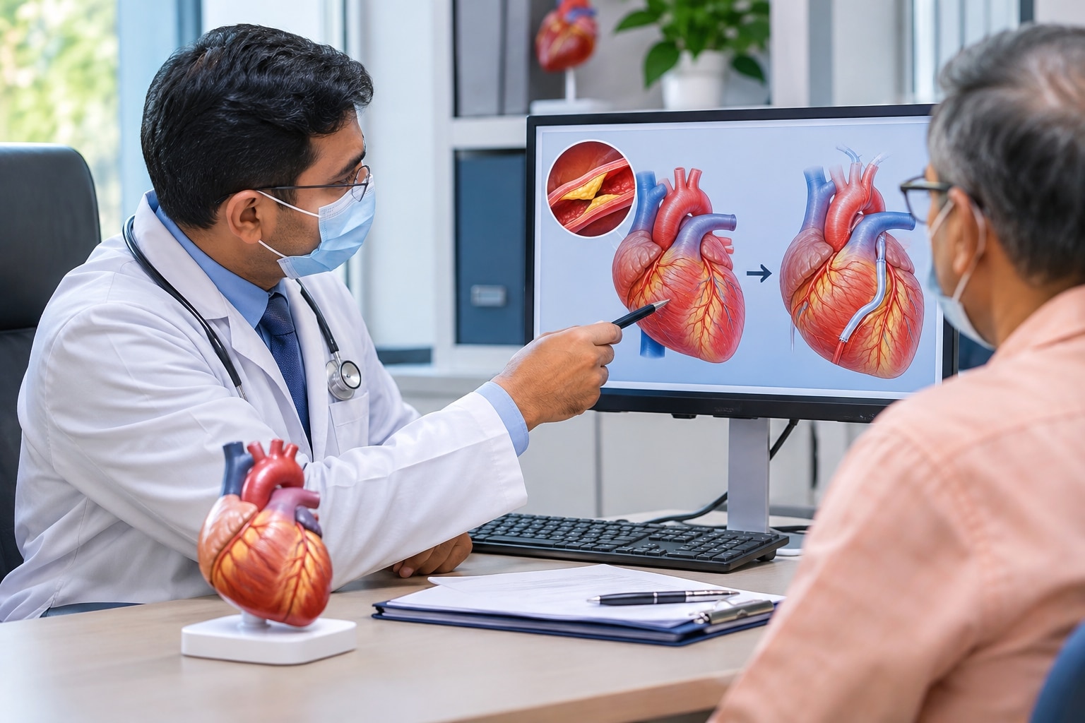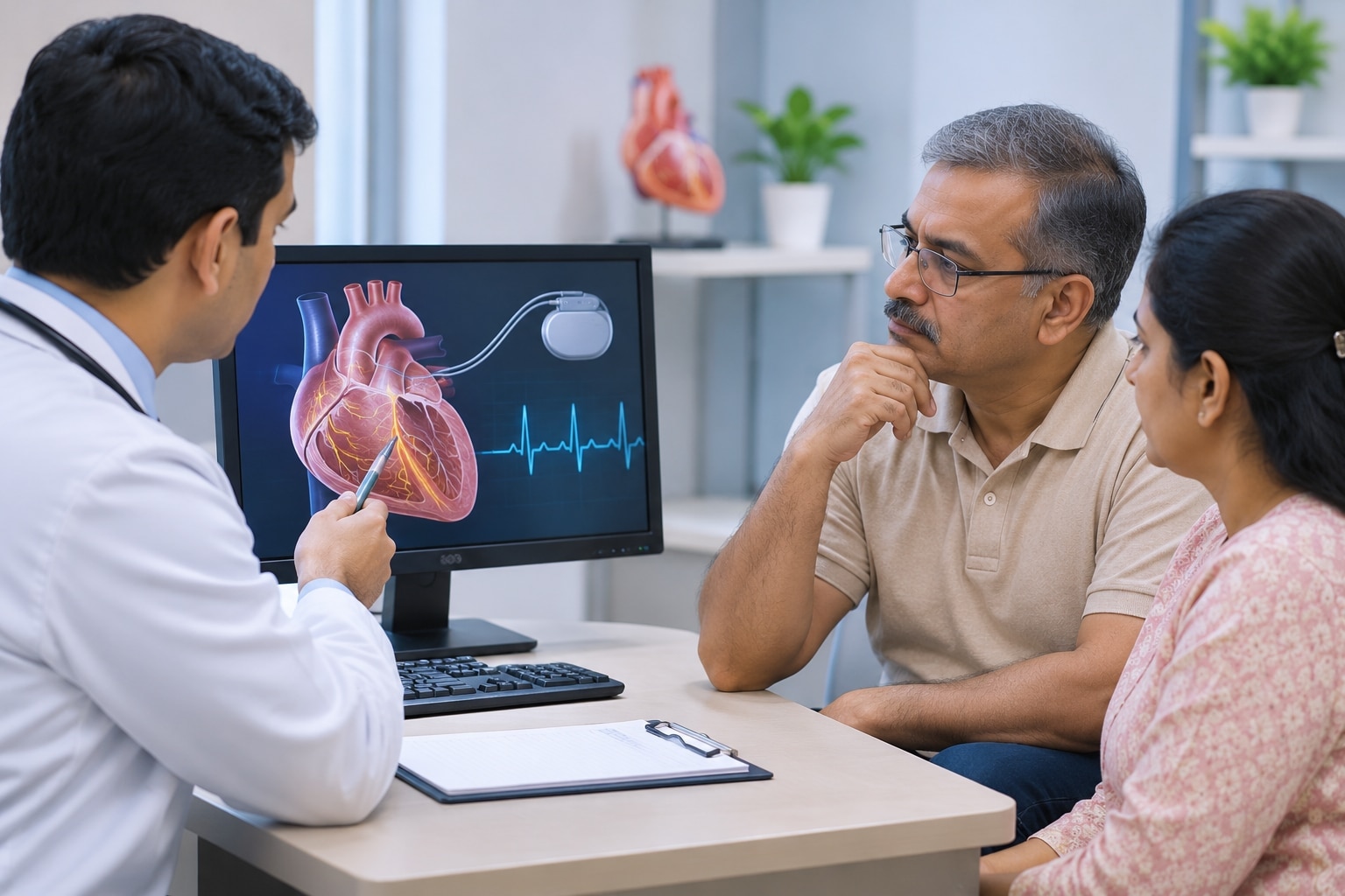
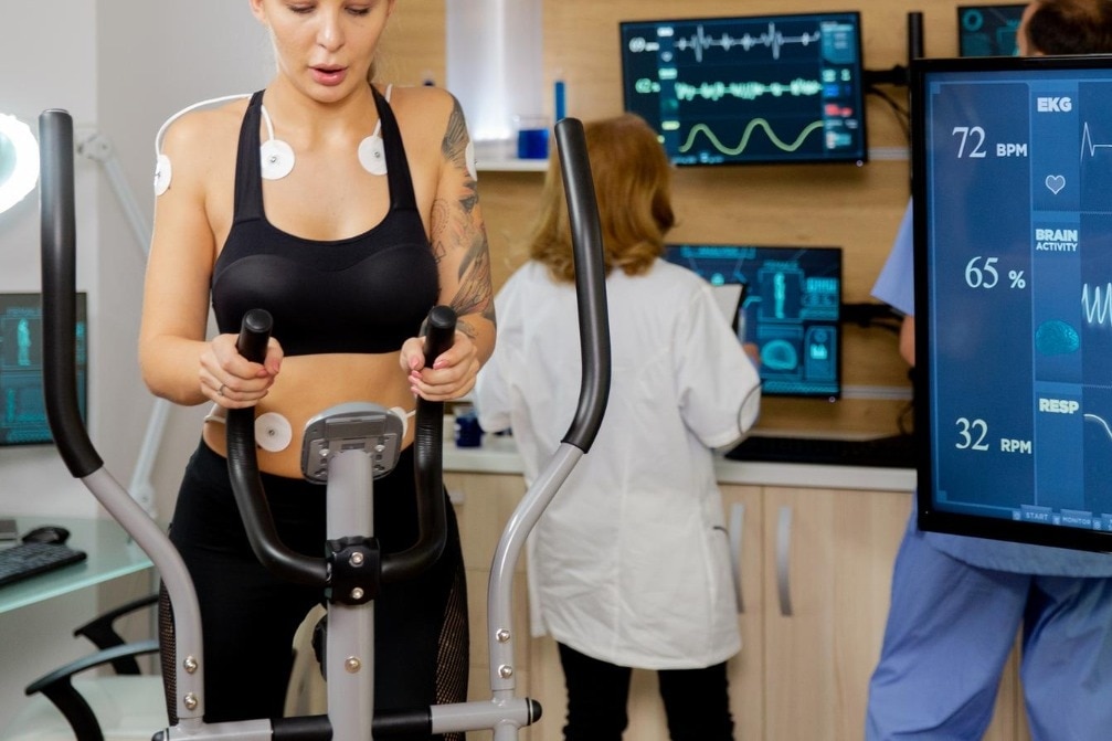
Before you walk on a treadmill, it helps to know which parts of heart function the test measures: heart rate, rhythm, blood pressure, exercise capacity and symptoms. You will also learn how to prepare, when exercise testing needs medical review, how to read the main report findings and which alternative test may answer a different cardiac question.
Key takeaways
- Review resting ECG, heart rate and blood pressure before exercise.
- Wear comfortable clothes and shoes suitable for walking.
- Postpone the test for acute illness or symptoms needing urgent care.
- Match the test to your question; TMT does not detect every heart problem.
What heart function is assessed before you start walking?
Before you start walking, a TreadmillStressTest records your resting heart rate, rhythm, blood pressure and baseline ECG. These readings show how your heart is functioning at rest and provide the comparison point for changes during exercise.
The assessment then follows the heart’s response to increasing workload:
- Heart rate: the monitor tracks how quickly your pulse rises and whether it reaches an appropriate level for the workload.
- Rhythm: continuous ECG monitoring looks for irregular beats or dangerous arrhythmias at rest, during exertion and in recovery.
- Blood pressure: staff measure the pressure response as exercise becomes harder. A poor rise, a significant fall or severe hypertension can require the test to stop.
- Exercise capacity: walking time, treadmill speed, incline and achieved workload show how much exertion you tolerate; this is not a general fitness score.
- Symptoms: report chest discomfort, unusual breathlessness, dizziness, leg fatigue or near-fainting immediately. Staff also assess how quickly symptoms, heart rate, blood pressure and ECG findings settle after walking.
A TMT is an exercise-provocation test, not a direct image of the coronary arteries. An abnormal finding does not automatically prove a blockage or mean angiography or a stent is needed. Your broader CardiacAssessment combines the result with age, symptoms, risk factors and the resting ECG.
What happens during a TMT appointment?
A TreadmillStressTest appointment usually follows this sequence:
1. Staff record your medical details, resting ECG and blood pressure before exercise. They explain the stop signal and ask about symptoms you already feel.
2. You remove clothing from the upper chest as needed, and staff place adhesive electrodes on prepared skin. Wires connect them to continuous ECG monitoring; a cuff remains on your arm for repeated blood-pressure readings.
3. You begin walking slowly on a level treadmill. In the standard Bruce protocol, speed and incline increase every three minutes, making each stage harder. A modified Bruce protocol starts with gentler stages for people with limited exercise capacity.
4. The staff watch your ECG, heart rate, blood pressure, walking pattern and symptoms throughout. Report chest pressure, unusual breathlessness, leg pain, dizziness, palpitations or weakness immediately.
5. Ten minutes is not a standard workload. Ten minutes on a gentle modified stage is very different from ten minutes near the upper Bruce stages, so the report records the protocol, completed stage and workload.
6. The test can feel physically demanding because both speed and incline rise. Stop when you cannot continue safely; staff do not expect you to force yourself to a predetermined duration.
7. Staff may stop the test before you reach a target heart rate for significant chest pain, severe breathlessness, near-fainting, dangerous rhythm changes, a major blood-pressure fall or rise, poor perfusion, or marked ECG changes.
8. You slow down or stop, then remain connected for recovery ECG and blood-pressure checks. Recovery symptoms and readings are part of the cardiac assessment.
How should you prepare, and when should you postpone the test?
Wear comfortable walking shoes and loose clothing for a TMTInErandwane appointment. Arrive 15 minutes early so staff can confirm your history, record baseline measurements and place electrodes without rushing.
1. Eat a light meal, or follow the clinic’s fasting instruction; avoid a heavy meal shortly before exercise. Do not smoke before the test. Ask whether you need to restrict caffeine: it is not routinely required for an ordinary exercise ECG, but it matters for some pharmacological or imaging stress tests.
2. Bring your prescription list and report beta-blockers, calcium-channel blockers, nitrates and every other medicine. Do not stop a beta-blocker or any prescribed drug yourself. These medicines can blunt your heart-rate response, but withholding them can be unsafe and can change what the test is intended to measure.
The ordering clinician must tell you whether to continue each medicine.
3. Tell staff about previous heart attack, angina, heart failure, valve disease, rhythm problems, fainting, high blood pressure, diabetes, recent procedures and any change in exercise tolerance.
Postpone routine exercise testing and seek medical review for chest pain at rest, severe breathlessness, fainting or near-fainting, an acute illness, uncontrolled blood pressure or a racing or irregular heartbeat with symptoms.
Recent heart attack, unstable angina, decompensated heart failure, acute myocarditis or pericarditis, pulmonary embolism, severe symptomatic aortic stenosis and uncontrolled arrhythmia require clinician clearance before proceeding. Stop and report symptoms during the test; reaching a target heart rate is not the only reason staff may end it.
How do you understand the results and their limits?
Exercise duration and achieved workload show how much exertion your heart tolerated. Workload is often reported in METs; 1 MET equals approximately 3.5 mL of oxygen per kilogram per minute. Reaching the target heart rate helps assess the test, but it is not the only measure of success.
The report also records blood-pressure response, ECG changes such as ST-segment depression, symptoms, arrhythmias and recovery after walking stops. A systolic pressure that fails to rise, falls, or becomes severely high changes the level of concern.
An abnormal TreadmillStressTest does not prove that a particular coronary artery is blocked. It may reflect reduced blood flow, but it can also require confirmation with another test; angiography or a stent is not automatic.
A negative result lowers concern under the right conditions but cannot exclude coronary disease or non-cardiac causes of chest pain, such as reflux, muscle pain or rhythm disorders.
Interpretation becomes difficult when the resting ECG already has left-bundle-branch block, ventricular pacing, left-ventricular hypertrophy with strain, pre-excitation, digitalis-related changes or marked ST abnormalities. The clinician must read the result as part of your CardiacAssessment, alongside symptoms, age, sex, risk factors, resting ECG, exercise capacity and recovery.
A symptom-free test with limited exercise capacity is not equivalent to a symptom-free test reaching a strong workload.
Which cardiac test answers the question you need answered?
Choose the test by the question you need answered, not by the test name or locality. A TMT is useful for exercise-related ECG changes only when you can exercise adequately and your resting ECG is interpretable.
| Test | Main question answered | Best fit |
|---|---|---|
| TMT | Do exertion, symptoms and ECG change during exercise? | Exercise-related ECG assessment |
| Resting ECG | What is the heart’s electrical pattern at rest? | Rhythm, conduction or baseline changes |
| Stress echocardiogram | Does heart muscle movement change under stress? | Ischaemia when exercise-ECG reading is limited |
| Nuclear stress test | Does blood flow to heart muscle fall during stress? | Perfusion assessment |
| Holter monitor | Does an intermittent rhythm problem occur during daily life? | Palpitations, dizziness or suspected arrhythmia |
| 2D echo | What do the chambers, valves and pumping function look like? | Structural and valve assessment |
A TMT can be the wrong first test if you cannot walk enough, have left-bundle-branch block, ventricular pacing or major baseline ST abnormalities. Your broader CardiacAssessment may instead need stress imaging or coronary CT angiography.
For anyone comparing TMTInErandwane facilities, check that they provide:
- Continuous ECG and blood-pressure monitoring
- Staff trained to recognise warning signs and stop exercise safely
- Emergency equipment immediately available
- Clinician interpretation, not only a printed tracing
- A documented plan for abnormal or inconclusive findings
Cardiologist in Pune can help match the test to your symptoms, exercise ability and ECG interpretability rather than treating TMT as suitable for everyone.
Related service
TMT (Stress Test) in Erandwane, Pune TMT (Stress Test) in Erandwane, Pune A Treadmill Test (TMT) is a non-invasive cardiac stress test that evaluates how your heart responds to physical activity. View service → |
Frequently asked questions
What heart function is assessed before starting a TMT?
The clinician records your resting heart rate, rhythm, blood pressure and baseline ECG before you walk. These readings provide the comparison point for exercise-related changes.
What happens during a TMT appointment?
The team attaches ECG electrodes, measures blood pressure, records baseline readings and increases treadmill speed or incline in stages while monitoring your symptoms and heart response.
How should you prepare for a TMT, and when should you postpone it?
Wear comfortable walking shoes and clothing, follow the clinic’s instructions about food and medicines, and tell the team about your health conditions. Postpone the test when you have an acute illness or new symptoms until a clinician advises you.
What do TMT results show, and what are their limits?
Results compare your symptoms, ECG, heart rate and blood-pressure response during exercise with your baseline. A TMT cannot answer every cardiac question, so your clinician may recommend another test.
Which cardiac test answers the question you need answered?
A TMT assesses the heart’s response to exercise. An echocardiogram evaluates heart structure and pumping, while other tests address rhythm, coronary arteries or symptoms that occur at rest.
Keywords