A coronary artery can look only moderately narrowed on an angiogram while plaque, calcium, or an underexpanded stent remains hidden from that two-dimensional view. By following the catheter’s path, understanding how echoes become images, and learning which measurements matter, you will be able to see what IVUS adds and when another test is needed.
Key takeaways
- IVUS uses sound waves inside a coronary artery during cardiac catheterization.
- Ask how IVUS measurements such as lumen area affect the treatment plan.
- An angiogram shows a contrast outline; IVUS shows the artery wall and plaque.
- Use coronary CT, cardiac MRI, or another test when IVUS cannot answer the question.
What happens during an IVUS examination inside a coronary artery
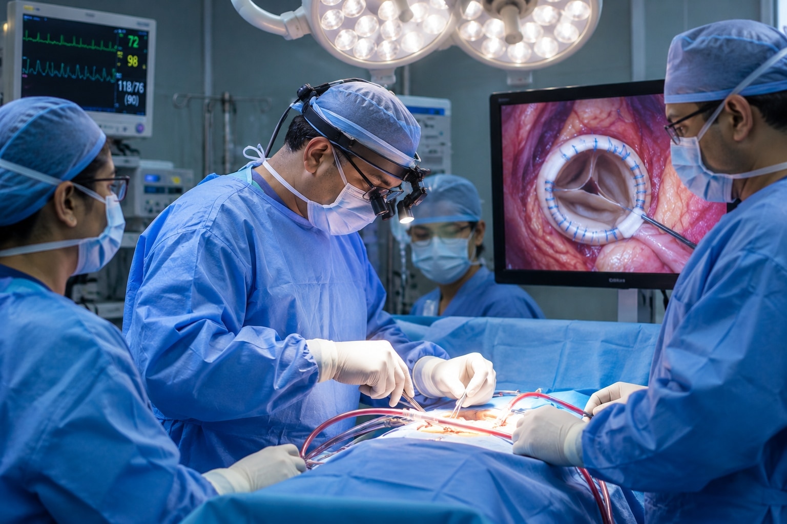
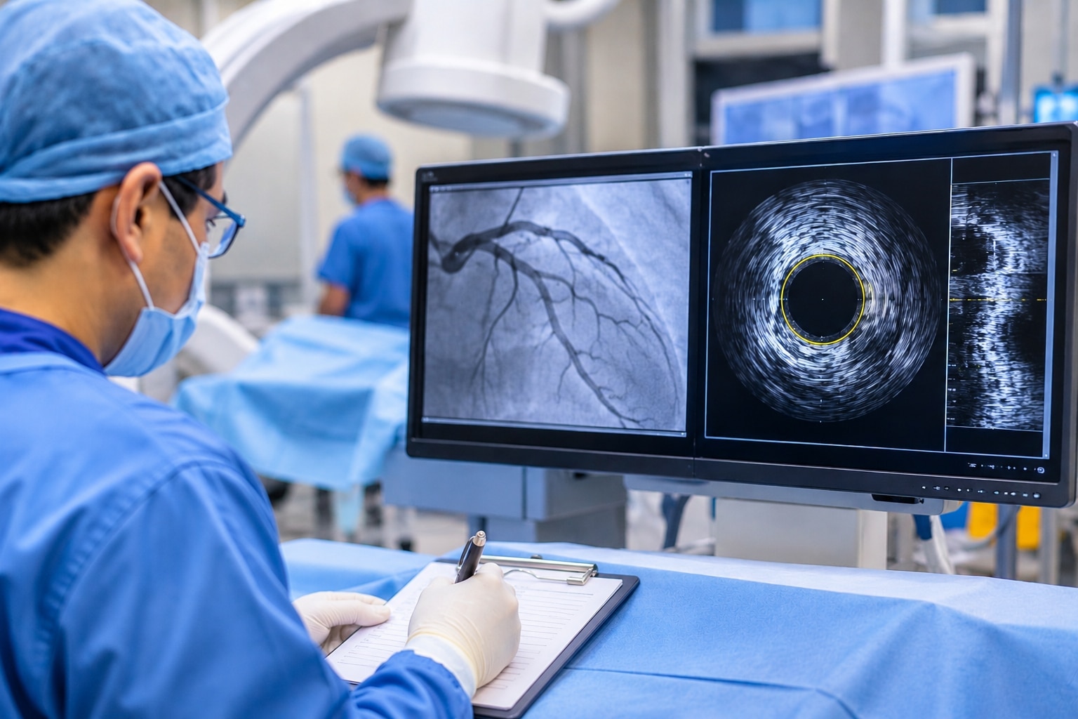
An IVUSProcedure takes place in a catheterization laboratory during cardiac catheterization, not in a standard ultrasound room. You lie on an examination table while staff monitor your heart rhythm, blood pressure, oxygen level and symptoms. IVUS itself uses sound waves, not ionizing radiation; the surrounding procedure can use X-ray fluoroscopy and contrast dye.
1. The team cleans and numbs the skin with a local anaesthetic, then enters an artery through the wrist (radial access) or groin (femoral access). You remain awake, although medication can help you relax.
2. The operator guides a thin sheath into the artery and passes a guiding catheter toward the heart. Contrast injections outline the coronary arteries on X-ray images, confirming catheter position and showing the target vessel.
3. A guidewire crosses the area being examined. The catheter-mounted IVUS probe travels over this wire into the coronary artery, usually beyond the region of interest.
4. The operator records images while withdrawing the probe at a motorized, measured speed or by hand. This pullback shows the lumen, plaque and vessel wall along the artery; it is the imaging sequence itself, not a separate skin incision.
5. The operator removes the imaging catheter, guidewire and guiding catheter. Staff apply firm pressure or a wrist compression device at the access site, then observe you for bleeding, pain, rhythm changes and circulation problems during recovery.
IVUS is invasive because instruments enter an artery and the coronary circulation. It is not a general screening test for healthy people; risks include bleeding, contrast reactions, vessel injury, spasm, clot and arrhythmia.
How ultrasound echoes become a cross-sectional coronary image
A miniature transducer on the catheter emits high-frequency sound waves, then records echoes returning from blood, plaque, calcium, the vessel wall and stent structures. The system converts the timing and strength of each echo into a circular cross-sectional image, with brighter signals marking stronger acoustic interfaces.
IntravascularUltrasound therefore shows what surrounds the open channel, not just the channel itself.
Image quality depends on how the catheter and sound beam interact with the artery:
- An off-centre catheter distorts apparent vessel diameter and plaque thickness.
- The guidewire creates a narrow shadow that can hide nearby tissue.
- Blood speckles or trapped air can produce noise, dropout or false boundaries.
- An uneven manual pullback changes the apparent length and spacing of a lesion.
- Heavy calcium appears bright but blocks sound, creating acoustic shadowing behind it; missing detail there does not prove healthy tissue.
The lumen is the blood-filled space inside the artery. The external elastic membrane, or the best visible outer vessel boundary, encloses the artery’s full size, including plaque within its wall.
Comparing these boundaries reveals plaque burden and positive or negative remodelling that a lumen-only view can miss: a vessel may expand outward while preserving a seemingly acceptable lumen. That distinction gives CoronaryArteryImaging greater structural detail than an outline alone.
Which measurements turn an IVUS image into a treatment decision
An IVUSProcedure turns a blockage percentage into measurements of the lumen, vessel, plaque, and stent—not just a picture. IntravascularUltrasound records the minimum lumen area (MLA), vessel diameter, reference vessel diameter, plaque burden, calcium distribution, and whether the artery has positive remodelling (enlargement) or negative remodelling (constriction).
| Measurement | What it shows | Treatment relevance |
|---|---|---|
| Minimum lumen area | Narrowest open area | Helps judge severity alongside anatomy and physiology |
| Plaque burden | Plaque as a proportion of vessel area | Guides lesion preparation and stent coverage |
| Calcium distribution | Arc, thickness, and length of calcium | Identifies need for calcium modification |
| Reference vessel diameter | Suitable healthy segment for comparison | Supports stent sizing and expansion targets |
| Vessel boundary | Full artery size beyond the lumen | Reveals remodelling hidden by a preserved lumen |
| Peri-stent tissue | Tissue, thrombus, or restenosis around or inside struts | Explains failure and guides further treatment |
After implantation, operators check the minimum stent area and expansion against an appropriate reference segment. They also look for:
- Incomplete apposition, where struts fail to touch the vessel wall
- Edge dissection or disease just beyond the stent
- Geographic miss, where diseased tissue lies outside the treated segment
- Tissue or thrombus protrusion through the struts
- Restenosis inside the stent
No single minimum-area threshold fits every vessel. The often-quoted 4 mm² is not a universal obstruction cutoff; the 6 mm² left-main deferral threshold applies to a specific setting. Interpret either value with vessel anatomy, symptoms, angiography, and physiology.
Why the angiogram and IVUS may tell different stories
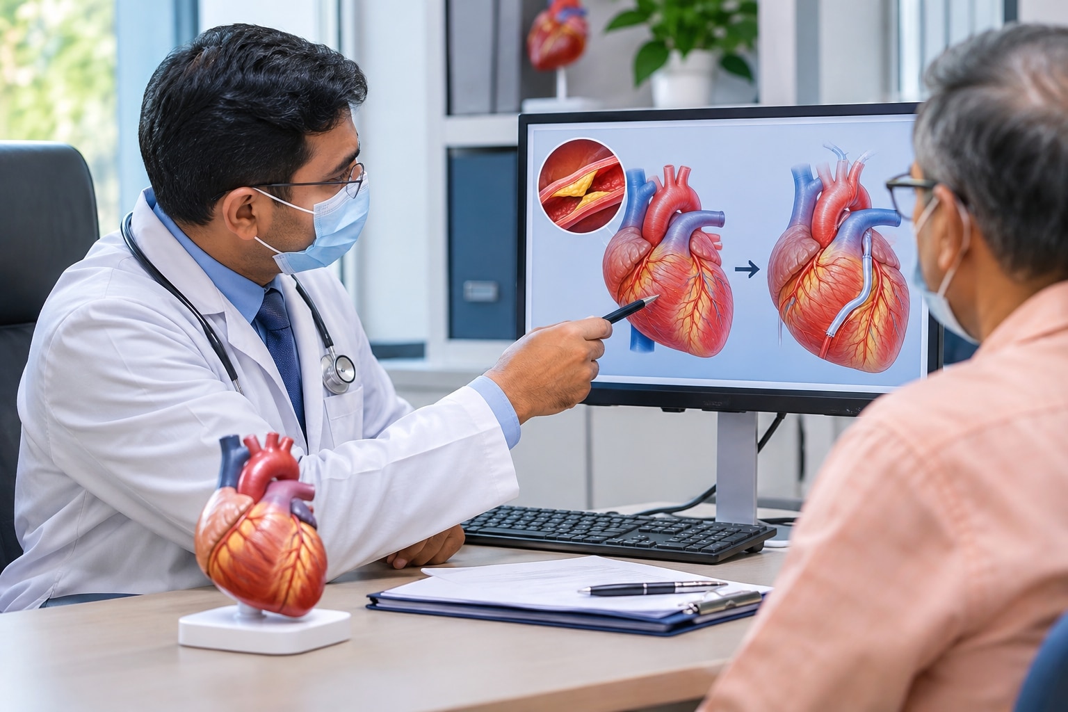
A two-dimensional contrast outline can underestimate or overestimate coronary disease because it shows the lumen from one viewing angle, not the artery’s full three-dimensional wall. Foreshortening makes a narrowing look shorter; vessel overlap can hide its tightest point.
Eccentric plaque can leave one side of the lumen looking wide, while diffuse narrowing lacks a normal segment for comparison. Plaque can also sit behind an apparently preserved lumen, masking substantial disease.
Angiography | IVUS What it shows | Contrast-filled lumen outline | Lumen, vessel wall, plaque, calcium and stent Common blind spot | Overlap, foreshortening and eccentric disease | Acoustic shadow behind heavy calcium Main measurement | Apparent narrowing in projection | Cross-sectional lumen area, vessel size and plaque burden
A cardiologist may add IVUS when the angiogram shows:
- An ambiguous lesion or possible left-main disease
- Heavily calcified plaque
- Long or diffuse narrowing
- Recurrent narrowing inside a stent
- An unexpectedly poor stent result
This CoronaryArteryImaging approach creates value when the findings change treatment: plaque preparation, stent diameter or length, landing position, or post-dilation. Recording a more detailed image without acting on underexpansion, malapposition, edge dissection or geographic miss adds little.
During an IVUSProcedure, the cross-sectional view can explain why a seemingly moderate lesion behaves as a severe one—or why a dramatic outline has less disease than expected. The image is useful when it changes the decision, not merely when it confirms what angiography already showed.
When IVUS is not enough and how to choose the next test
IVUS cannot by itself prove that a narrowing causes chest pain or determine the physiological significance of every borderline lesion. A 4 mm² minimum lumen area is not a universal obstruction cutoff; even the 6 mm² left-main deferral threshold applies to a specific setting, not every artery.
Choose the next test according to the unanswered question:
| Option | Best suited to | Limitation |
|---|---|---|
| IVUS | Vessel size, plaque burden, calcium, remodelling, and stent expansion | Cannot directly prove flow limitation; calcium and catheter position can distort measurements |
| FFR | Whether an intermediate narrowing reduces pressure and blood flow during hyperaemia | Requires pressure-wire measurement and medication-induced hyperaemia |
| iFR | Flow-related significance using a resting pressure measurement | Results still need clinical and anatomical context |
| OCT | Thin fibrous caps, tiny dissections, and clear stent-strut detail | Needs a rapid contrast or flush injection to clear blood and penetrates less deeply than IVUS |
IVUS usually needs less prolonged blood clearance and reaches deeper tissue. OCT’s sharper detail can expose findings that IVUS misses, while heavy calcium can create acoustic shadowing that limits either interpretation. Uneven pullback, guidewire shadow, blood or air artefacts, and incorrect reference segments also affect measurements.
Both tests are invasive and share risks including arterial bleeding, contrast reaction, coronary spasm, dissection, thrombosis, arrhythmia, and vessel injury. A cardiologist in Pune can decide whether invasive CoronaryArteryImaging is justified, then match the physiology or IntravascularUltrasound test to your symptoms, angiogram, anatomy, and treatment question—not use IVUS as a routine scan.
Related service
Intravascular Ultrasound (IVUS) in Erandwane, Pune Intravascular Ultrasound (IVUS) in Erandwane, Pune Intravascular Ultrasound (IVUS) is an advanced imaging test that captures detailed cross-sectional images of coronary arteries from inside the... View service → |
Frequently asked questions
What happens during an IVUS examination inside a coronary artery?
During cardiac catheterization, a doctor guides a thin catheter into a coronary artery and passes an IVUS probe through it. Staff monitor your heart rhythm, blood pressure, oxygen level, and symptoms. IVUS uses sound waves, while fluoroscopy and contrast dye may support the surrounding procedure.
How does IVUS create a cross-sectional coronary image?
The IVUS probe sends high-frequency sound waves into the artery and records returning echoes. Software converts differences in echo strength and timing into a circular cross-sectional image showing the lumen, plaque, calcium, and vessel wall.
Which IVUS measurements help guide treatment?
Doctors assess the minimum lumen area, reference vessel diameter, plaque burden, lesion length, and stent expansion. These measurements help determine whether a narrowing is significant and whether a stent is positioned and expanded properly.
Why can an angiogram and IVUS show different findings?
An angiogram displays a two-dimensional silhouette created by contrast flowing through the artery. IVUS examines the artery from inside, revealing plaque hidden within the vessel wall, remodeling, calcium, and the true lumen dimensions.
When is IVUS not enough, and what test comes next?
IVUS cannot answer every question. Doctors may add fractional flow reserve or instantaneous wave-free ratio to assess blood-flow limitation, coronary CT angiography for noninvasive anatomical imaging, or cardiac MRI when tissue function or scar is the concern.
Keywords